
Image result for pericardium Circulatory system, Cardiovascular system, Anatomy and physiology
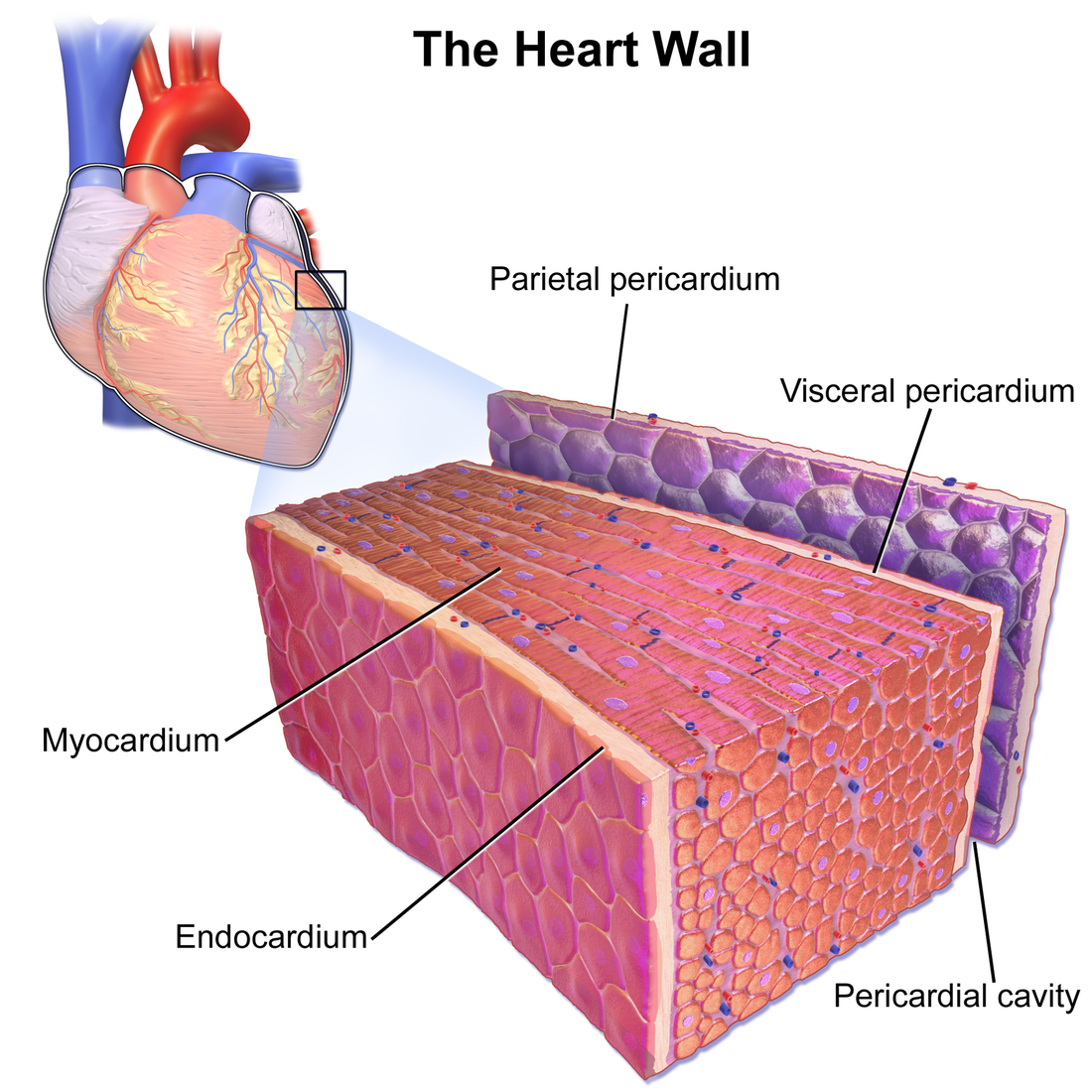
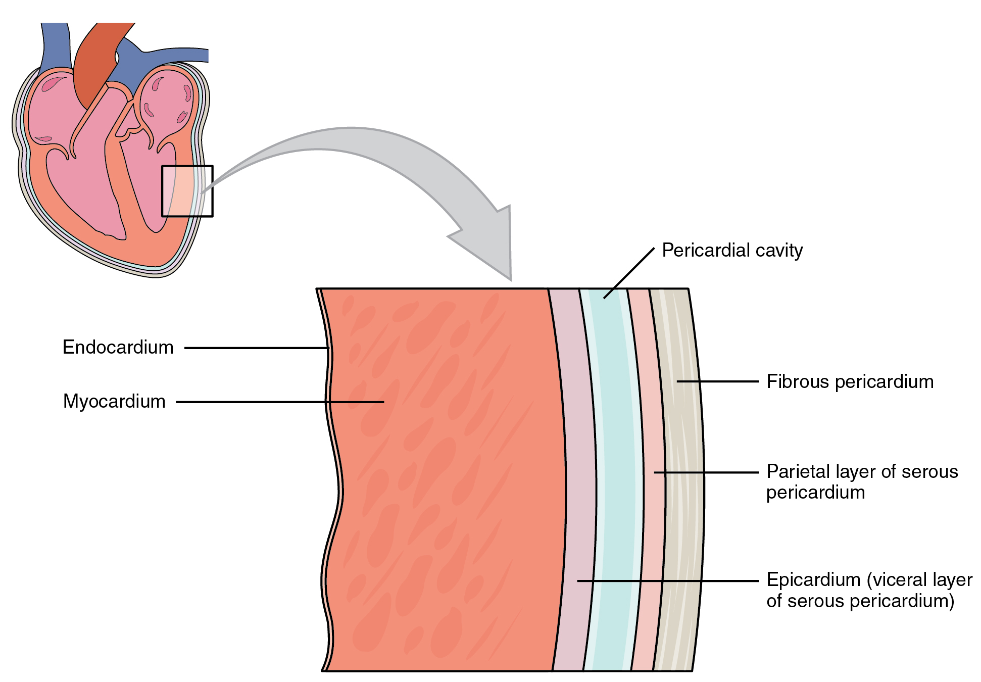
The epicardium is the outermost layer of the heart. It is actually the visceral layer of the serous pericardium, which adheres to the myocardium of the heart. Histologically, it is made of mesothelial cells, the same as the parietal pericardium. Below the mesothelial cells is a layer of adipose and connective tissue that binds the epicardium to.
Pericardial Fluid Urinalysis and Body Fluids
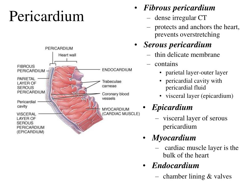
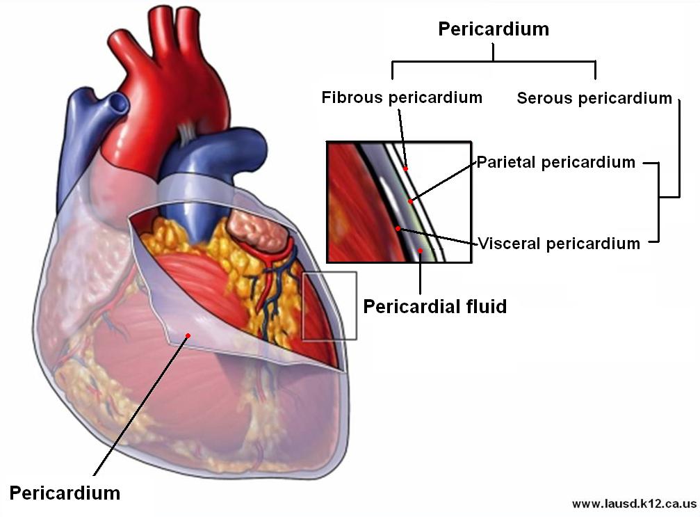
The pericardium is a fluid-filled doubled-walled membrane sac that surrounds the heart. The fluid is separated by two layers, the fibrous and serous pericardium.[1] The fibrous pericardium is the outer layer and holds the heart in place and protect it from surrounding infections.[1] It is composed of thick connective tissue. The serous pericardium has two layers, the visceral and parietal layers.
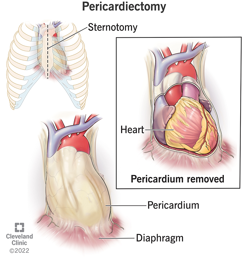
Pericardiectomy Details, Recovery and Outlook
In fact, each day, the average heart beats 100,000 times, pumping about 2,000 gallons (7,571 liters) of blood. Your heart is located between your lungs in the middle of your chest, behind and slightly to the left of your breastbone (sternum). A double-layered membrane called the pericardium surrounds your heart like a sac.

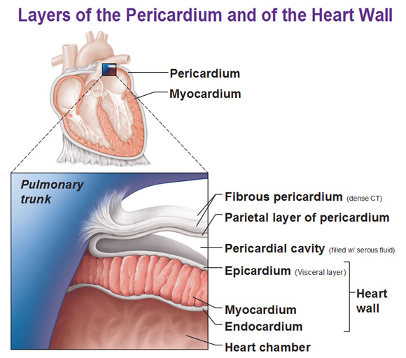
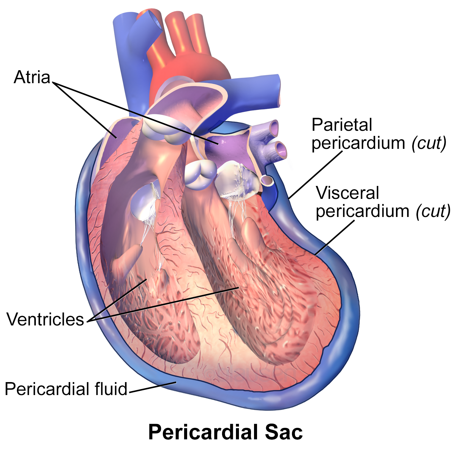
The pericardium is a doublewalled sac that encloses the heart. Between the visceral and
Rarely, a pericardial cyst can lead to heart failure.. Constrictive pericarditis is chronic inflammation of the pericardium, which is a sac-like membrane that surrounds the heart. READ MORE.
PPT Anesthesia with Cardiac Tamponade PowerPoint Presentation ID299640
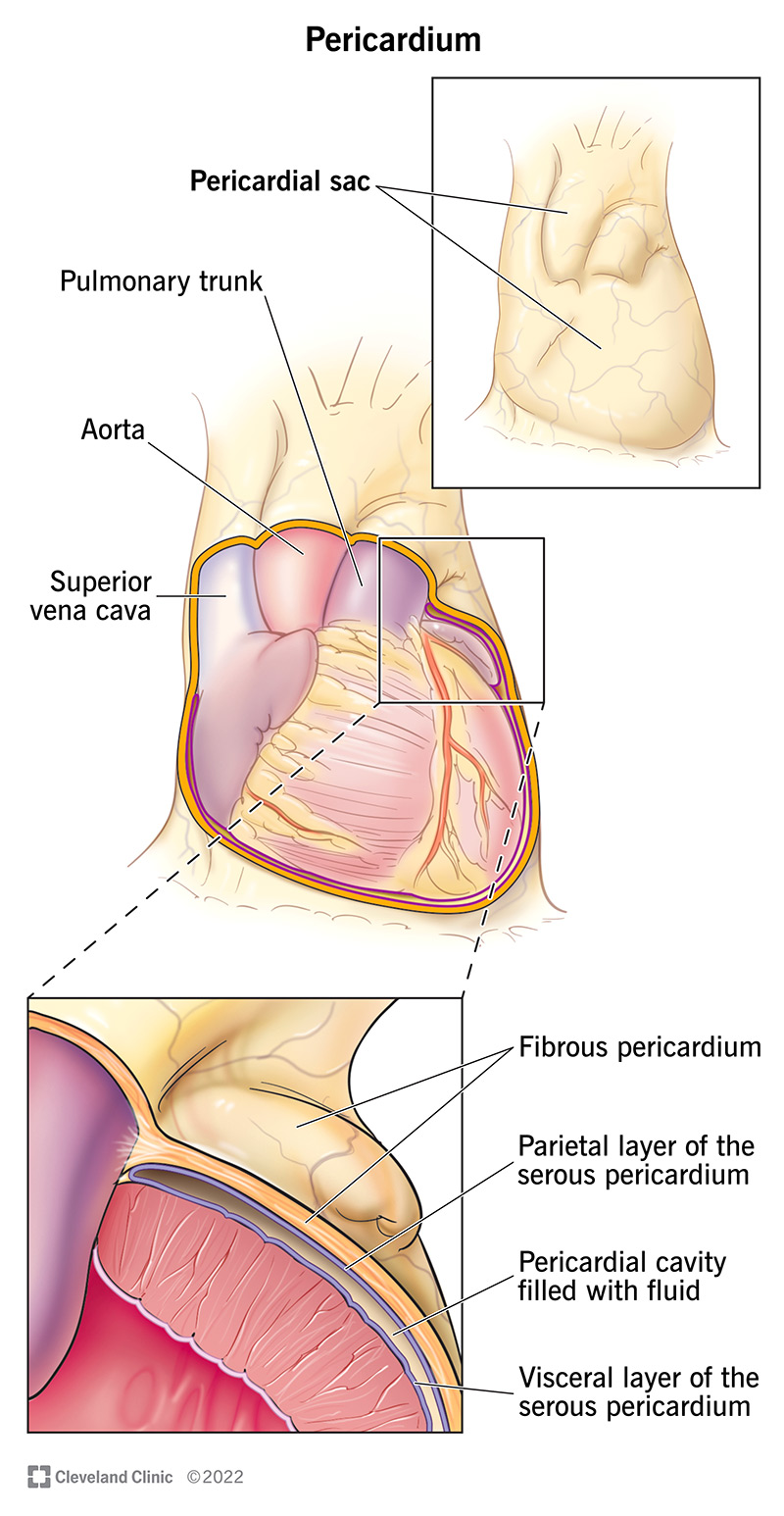
The pericardial membrane and the heart wall share the epicardium. The membrane that directly surrounds the heart and defines the pericardial cavity is called the pericardium or pericardial sac. It also surrounds the "roots" of the major vessels, or the areas of closest proximity to the heart. The pericardium, which literally translates as.
PPT The Cardiovascular System The Heart PowerPoint Presentation, free download ID312194
The pericardium is the thick, membranous, fluid-filled sac that surrounds the heart and the roots of the vessels that enter and leave this vital organ, functioning as a protective membrane. The pericardium is one of the mesothelium tissues of the thoracic cavity, along with the pleura which cover the lungs. The pericardium is composed of two.
Heart Anatomy · Anatomy and Physiology
The heart resides within the pericardial sac and is located in the mediastinal space within the thoracic cavity. The pericardial sac consists of two fused layers: an outer fibrous layer and an inner parietal pericardial serous membrane. Between the pericardial sac and the heart is the pericardial cavity, which is filled with lubricating serous.
Pericardium Function and Anatomy
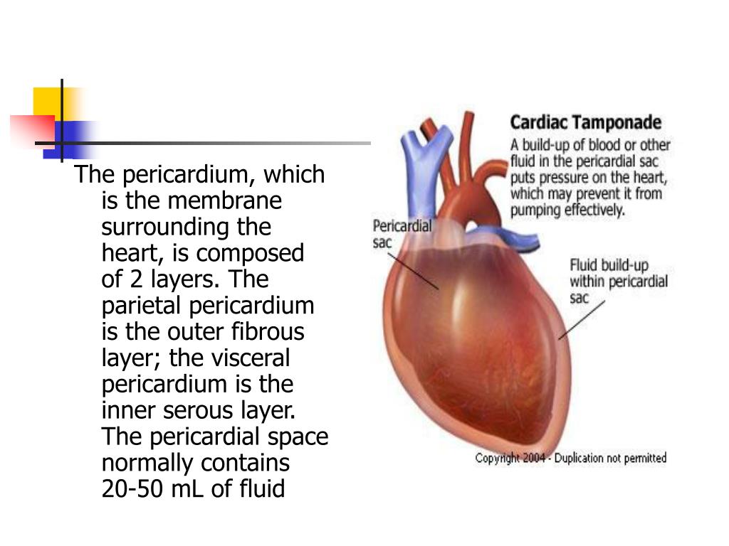
The pericardial cavity lies between the visceral pericardium and the parietal pericardium. This cavity is filled with pericardial fluid which serves as a shock absorber by reducing friction between the pericardial membranes. There are two pericardial sinuses that pass through the pericardial cavity. A sinus is a passageway or channel.

Pericardium Definition & Function Video & Lesson Transcript
The pericardial membrane and the heart wall share the epicardium. Figure 9.5: Pericardial Membranes and Layers of the Heart Wall. Surface Features of the Heart. Inside the pericardium, the surface features of the heart are visible, including the four chambers. There is a superficial leaf-like extension of the atria near the superior surface of.
Layers of the Pericardium, Heart Wall and Spiral Arrangement
If the heart is the fun, interesting inside bit of an orange, the pericardium could be compared to the peel around it.Like peel, it can seem vaguely unexciting - that is until you learn some of its very important (appeeling. ahem.) physiological functions 1. In scientific terms, the pericardium is a fibro-serous, fluid-filled sack that surrounds the muscular body of the heart and the roots.
Print The Heart flashcards Easy Notecards
Your pericardium is a protective, fluid-filled sac that surrounds your heart and helps it function properly. Your pericardium also covers the roots of your major blood vessels as they extend from your heart. These are known as your "great vessels," and they include your: Aorta. Main pulmonary artery. Pulmonary veins.
/pericardium-57a8a12e5f9b58974a2b4fb7.jpg)
Pericardium—Anatomy and Function
The pericardium is a membrane, or sac, that surrounds your heart. It holds the heart in place and helps it work properly. Problems with the pericardium include: Pericarditis - an inflammation of the sac. It can be from a virus or other infection, a heart attack, heart surgery, other medical conditions, injuries, and certain medicines.

Location of the heart Human Cardiovascular System
The pericardium is a fibrous sac that encloses the heart and great vessels. It keeps the heart in a stable location in the mediastinum, facilitates its movements, and separates it from the lungs and other mediastinal structures. It also supports physiological cardiac function.[1][2][3]
PPT Pericardium & Heart PowerPoint Presentation, free download ID4956744
When you have pericarditis, the membrane around your heart is red and swollen, like the skin around a cut that becomes inflamed. The pericardium is a thin, two-layered, fluid-filled sac that covers the outer surface of your heart. It provides lubrication for your heart, shields it from infection and malignancy, and contains your heart in your.
Pericardium The Heart Protector Dr. Elizabeth Cox, ND, LAc
The pericardium ( pl.: pericardia ), also called pericardial sac, is a double-walled sac containing the heart and the roots of the great vessels. [1] It has two layers, an outer layer made of strong inelastic connective tissue ( fibrous pericardium ), and an inner layer made of serous membrane ( serous pericardium ).

membrane called pericardium peri around cardium greek this actually image Double Layered
The pericardium is a fluid-filled sac that encases the muscular body of the heart and the roots of the great vessels (including the aorta, pulmonary trunk, pulmonary veins, and the inferior and superior vena cavae ). This fibroserous sac is comprised of a serous membrane supported by a firm layer of fibrous tissue.