
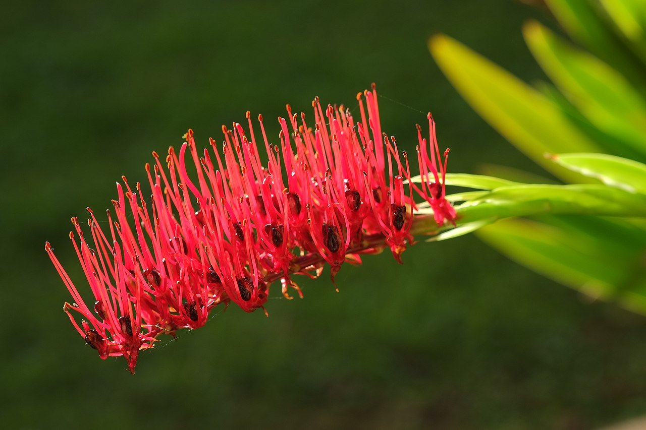
Pin on NZ Native grasses
The question as to how many chromonemata may actually be seen in large somatic plant or animal chromosomes has been summarized by Sharp (1934), later by Kaufmann (1936) and still later by Geitler (1938a). Darlington (1937a) still maintains that the chro-mosome does not split until the division commences during which half-chromosomes separate.

Xeronema callistemon Native garden, Trees to plant, Plants
14 Similar questions Q. Chromonemata are embedded in a Q. Each chromonemata contains Q. Chromonemata start associating into bivalent chromosomes during Q. A gaint chromosome with a number of chromonemata is Q. During synapsis the number of thread (Chromonemata) in each chromosome is: View More Introduction BIOLOGY Watch in App

Col SEM of giant chromosome from salivary gland Stock Image P657/0014 Science Photo Library
chro·mo·ne·ma·ta ( krō'mō-nē'mă, -ma-tă ), The coiled filament in which the genes are located, which extends the entire length of a chromosome and exhibits an intensely positive Feulgen test result for DNA. Synonym (s): chromatic fiber [chromo- + G. nēma, thread] Farlex Partner Medical Dictionary © Farlex 2012 chromonema (krō′mə-nē′mə)
your biology book here's what chromosomes really look like
Chromatin, chromosome, chromatid, chromonema, chromonemata and chromomere- these are sound very similar but are actually different things. These terms are ve.

PlantFiles Pictures Xeronema Species, Poor Knight's Lily (Xeronema callistemon) by mrporl
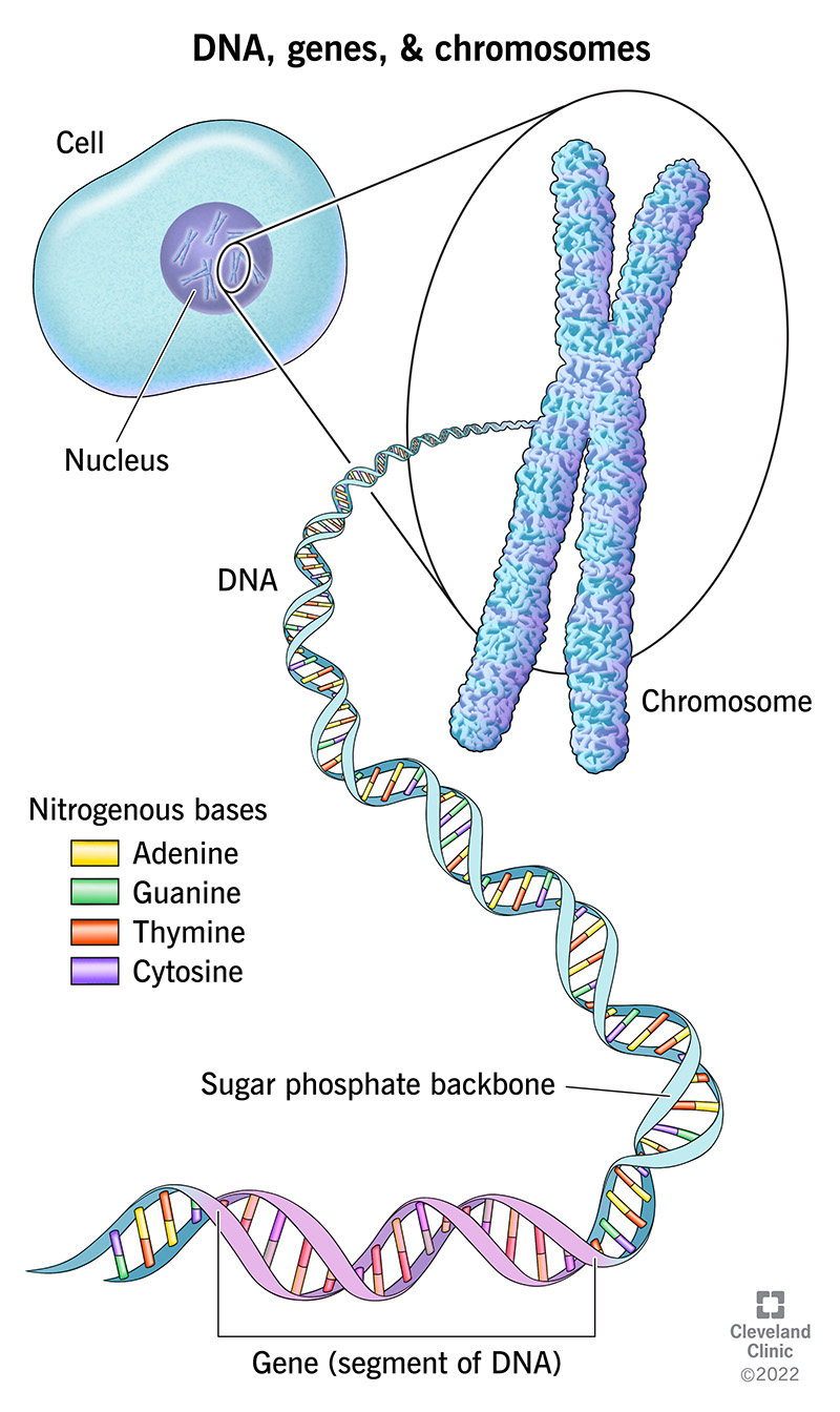
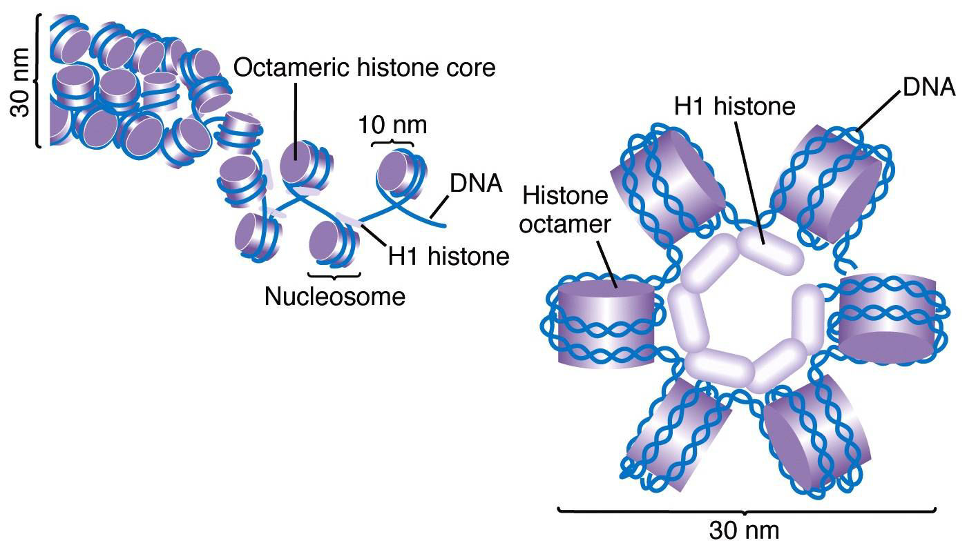
The following levels of DNP compaction in mitotic chromosomes are suggested: a 10-nm nucleosomal fibril, a 25-nm nucleomeric fibril, and the chromonema, a fibrous structure, about 100 nm in diameter, composed of chromomeres. Interphase nuclei also contain structures which are morphologically similar to the chromomeres of mitotic chromosomes.

Xeronema Callistemon, Poor Knights Lily Stock Image Image of shaped, xeronema 51617363
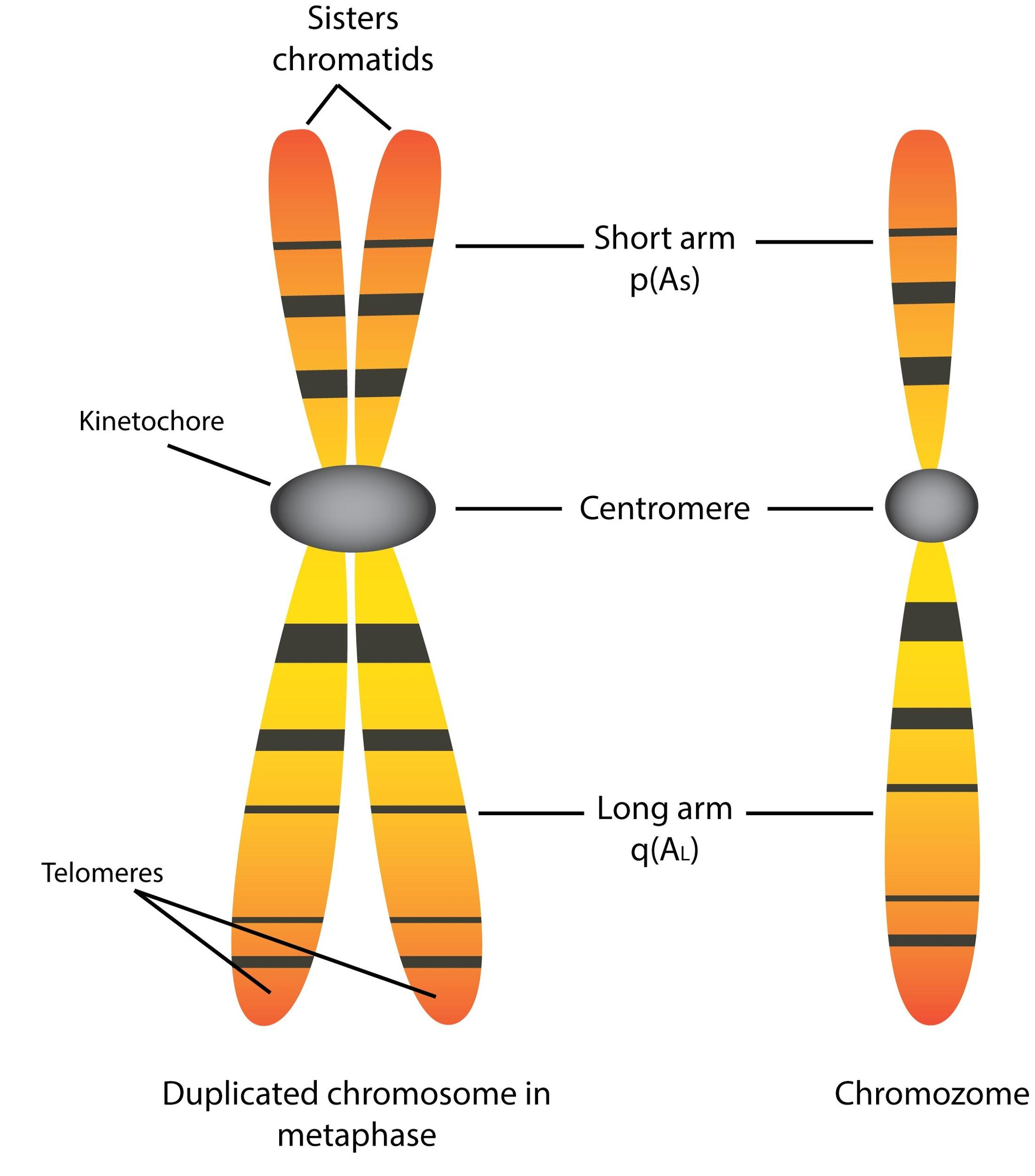
Chromonema was first of all observed by Baranetzky in 1880, in the pollen mother cell of Tradescantia, and was called chromonema (singular) by Vejdovsky in 1912. At metaphase each chromosome consists of two symmetrical structures, the chromatids, each of which contains a single DNA molecule.

How to Pronounce Chromonemata YouTube
The number of chromonemata is not fixed in each chromatid. It varies from 2 to 32 in number. During prophase, the chromosome becomes visible and filamentous called chromonemata. Cromonemata form gene bearing portion of the chromosomes. The bead-like appearance of chromatin material on chromonemata is called chromomeres.
Describe the structure of the chromosome with a suitable diagram.
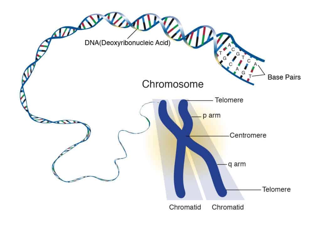
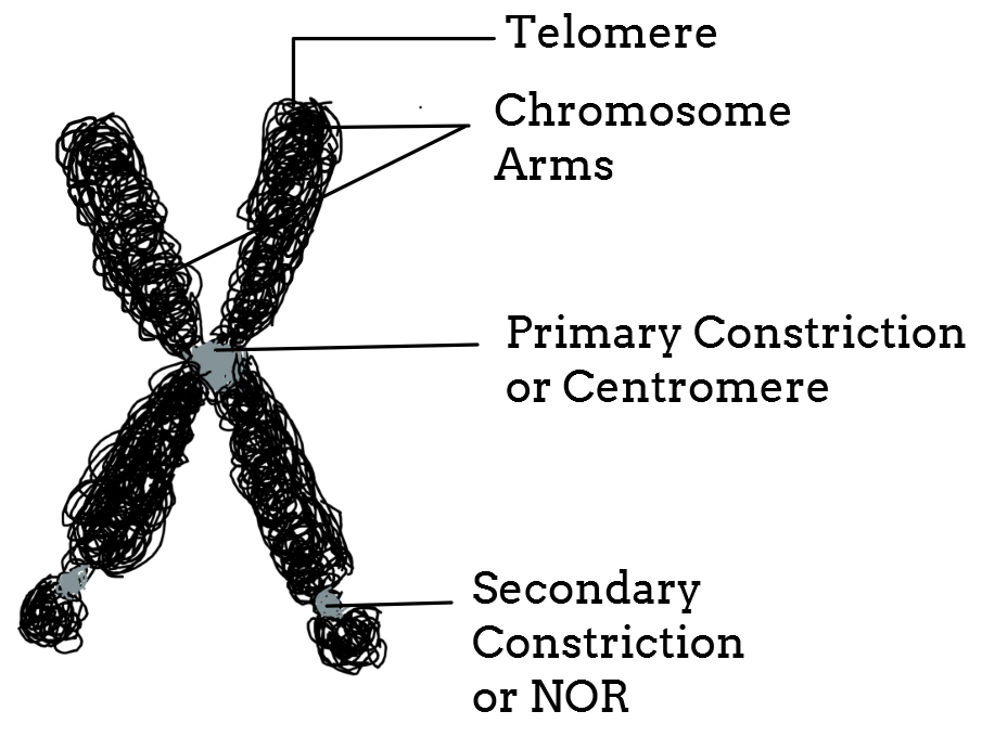
c. Chromonemata, d. Primary constriction, e. Secondary constriction, f. Satellite and. g. Telomere. a. Pellicle: It is the outer, thin but doubtful covering or sheath of the chromosome. b. Matrix: Matrix or ground substance of the chromosome is made up of proteins, small quantities of RNA and lipid.
Pasta Is Made Up Of Wholesale Shop, Save 61 jlcatj.gob.mx
chro·mo·ne·ma·ta ( krō'mō-nē'mă, -ma-tă ), The coiled filament in which the genes are located, which extends the entire length of a chromosome and exhibits an intensely positive Feulgen test result for DNA. Synonym (s): chromatic fiber [chromo- + G. nēma, thread] Farlex Partner Medical Dictionary © Farlex 2012 chromonema (krō′mə-nē′mə)

Figure 1 from The Hydration and Dehydration Phenomena in MitosisIV. The chromonemata as natural
chromonema Quick Reference (pl. chromonemata) cytological term for 1 all of the threads which make up the nuclear reticulum. 2 any of the smallest strands of DNA in a chromosome or chromatid. 3 a twisted chromatid thread within the chromosome. [.] From: chromonema in Oxford Dictionary of Biochemistry and Molecular Biology »

Chromosome Definition, Structure, Types and Function Biology Ideas
6 Main Parts of a Chromosome Article Shared by ADVERTISEMENTS: The following points highlight the six main parts of a chromosome. The parts are: 1. Pellicle and Matrix 2. Chromatids, Chromonema and Chromomeres 3. Centromeres 4. Secondary Constriction 5. Satellite 6. Telomere. Part # 1. Pellicle and Matrix:
What Is A Chromosome B Fa My XXX Hot Girl
What are Chromosomes? Structure of a Chromosome Pellicle Matrix Chromonemata Centromere Secondary Constriction or Nucleolar Organiser Telomeres Types of Chromosomes A. Autosomes and Sex Chromosomes B. On the Basis of Number of Centromeres C. On the Basis of Location of Centromere Prokaryotic Chromosomes Eukaryotic Chromosomes a. Nucleosomes
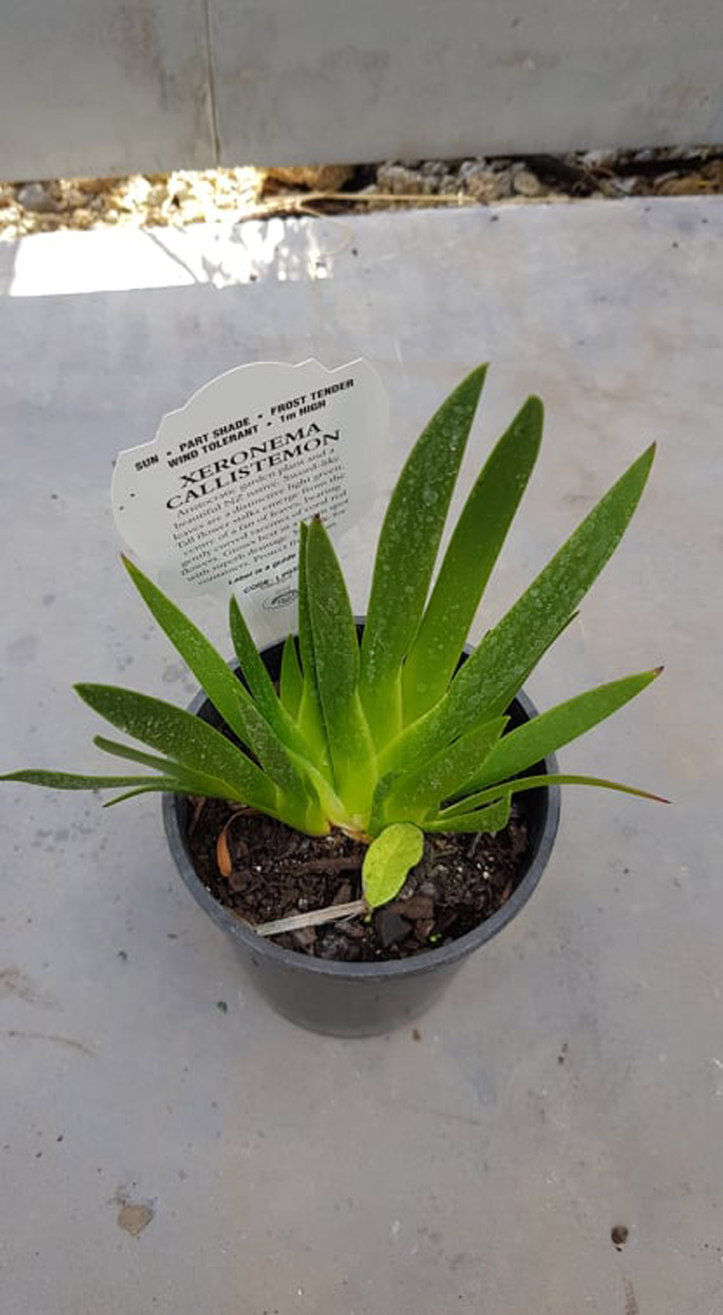
Xeronema Springvale Garden Centre
1 10nm fibres are seen only by electron microscopy,while chromonema fibres refer to chromatin fibres visible by light microscopy. See this paper. 10nm or 30nm fibres are thus much, much smaller than chromenema fibres. There are likely several layers of organization (like 100nm and 200nm fibres) before you get all the way up to chromenema fibres.
Chromosome structure hercwules
Chromonemata is the gene-bearing structure of a chromosome. Sometimes (in interphase), bead-like accumulations of chromatin material are visible along the chromosomes. These are termed as chromomeres. These are regions of tightly-packed DNA. Usually, the centromere lies within the primary constriction (thinner chromosomal segment).
Xeronema callistemon (Poor Knight's Lily) Kat's Garden
What is the Chromosome structure? How is DNA packaged into chromosomes and describe the structure of a chromosome? The general structure of somatic chromosomes can be studied best at the metaphase and anaphase of mitosis. Each comprises the following parts: Pellicle and Matrix Chromonemata (Chromatid during Metaphase) Chromomeres Centromere

Chromatin, chromosome, chromatid, chromonema, chromonemata,chromomere YouTube
The chromonema is the carrier of the genes. They also help in maintaining the proper structure of chromomere. The chromonema appears to be like a mass of coiled threads at the beginning of the cell division. The chromonema bears several knot or beaded structure which is called chromomere. These granules are present at regular intervals.